
Inside Rodin's Hands
dal 8/4/2014 al 2/8/2014
Segnalato da


8/4/2014
Inside Rodin's Hands
Cantor Arts Center, Stanford
Art, Technology, and Surgery. The exhibition allows visitors to experience Rodin's hands as Dr. Chang's students do in the seminar. It presents the modern surgeon's diagnostic process.
Stanford, CA— James Chang fell in love with the Rodin Sculpture Garden at Stanford when he was an undergraduate. Later, while training in plastic and reconstructive surgery at the Stanford University School of Medicine, he visited the Cantor Arts Center with his family on Thursday evenings, where they would enjoy dinner in the café and then the art. His fascination for the work of the famous French artist Auguste Rodin (1840–1917) grew as Dr. Chang saw signs in the sculptures of the medical conditions he was learning to identify. Now as a professor in Stanford’s medical school, he shows images of Rodin’s sculptures of hands to medical students and hand surgery trainees, asking them to make diagnoses and hoping the artworks will make learning more fun and memorable.
Dr. Chang’s enthusiasm for this art has also fueled his undergraduate seminar, “Surgical Anatomy of the Hand: From Rodin to Reconstruction” with students studying the hands that Rodin created and their perceived medical conditions aided by advanced technology. Three-dimensional scans of the sculptures combined with CT scans of patients’ bones, nerves and blood vessels create a new “augmented reality” that reveals the pathologies beneath the bronze and even allows students to perform virtual surgery.
This remarkable seminar inspired an exhibition that allows museum visitors to experience Rodin’s hands in much the same manner as Dr. Chang’s students. “Inside Rodin’s Hands: Art, Technology, and Surgery” opens at the Cantor April 9 and continues through August 3. The exhibition, unique in how it merges art and science, involved an unprecedented collaboration between four diverse groups at Stanford: the Cantor Arts Center, Dr. Chang and his students, the Division of Clinical Anatomy, and the Lane Medical Library.
“This exhibition brings together the best of Stanford with a cross-disciplinary set of contributors,” said Cantor Director Connie Wolf. “We were all inspired by Dr. Chang and his passion. Outstanding individuals from the anatomy department at the School of Medicine contributed unique, cutting-edge technology that is changing how surgery is taught throughout the world. At the Cantor, we offered our curatorial expertise, the renowned Rodin collection, and our commitment to interdisciplinary approaches to the arts. And the medical school’s Lane Library is lending important historical materials. The groups worked so well together, this is truly a project that could only have happened at Stanford.”
Dr. Chang cited the unprecedented collaboration for the success of the overall project, the seminar and the exhibition. He added, “I wanted to participate in this exhibition for the same reason I introduced Rodin into my seminar: to get students in the humanities excited about the sciences, and to get doctors to step out of the hospital to appreciate art. I have found that artists and surgeons appreciate human anatomy with equal passion.”
The Exhibition
The exhibition allows visitors to experience Rodin’s hands as Dr. Chang’s students do in the seminar. After an introduction to Rodin, his art, and his appreciation of the hand as an especially expressive part of the human body, the exhibition presents the modern surgeon’s diagnostic process. Rodin bronzes on display are accompanied by evaluations of the sculptures’ perceived conditions, written by Dr. Chang and his students. Photographs of patients’ hands with the same conditions—Dupuytrens contracture, Apert syndrome, Charcot-Marie-Tooth disease, ganglion cyst, thumb amputation, fracture and stiff joint— illustrate how surgeons correct the conditions.
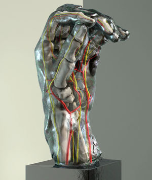
Visitors see what lies “beneath the skin” of the bronze hands—the underlying anatomy that Dr. Chang imagined when he encountered the sculptures at the Cantor—through the use of augmented reality. By rotating an iPad in an arc around three of Rodin’s hand sculptures, visitors can see computer-generated graphics of bones, nerves and blood vessels from varying angles. Dr. Chang uses this technology in his class because it gives his students a sense of the hand’s three-dimensional structure, an experience otherwise limited to time working on actual cadavers. In Dr. Chang’s class, students go on to perform virtual surgery on the hands, a teaching method likely to become increasingly prevalent in medical schools.
The creation of the computer-generated images for the Rodin-hands augmented reality required close cooperation between diverse Stanford medical groups. After hyper-detailed scans were made of the exterior of the bronzes, Dr. Paul Brown and his colleagues at Stanford’s Division of Clinical Anatomy put CT scans of the internal elements of some of Dr. Chang’s patients into the scans of the bronzes. (See “The Division of Clinical Anatomy,” below.)
An added feature of the exhibition puts the study of anatomy into historical context with medical texts published between the 16th and 19th centuries. Illustrations in these volumes, lent by the Lane Medical Library and Stanford’s Special Collection, help visitors see how earlier generations studied the anatomy of the hand. Labels written by Michael Bartolos (’14, M.F.A.), Kathleen Chang (Dr. Chang’s high-school age daughter) and Susan Chang (’15, Physics) explain the importance of the exquisitely detailed illustrations.
Videos on view depict virtual hand surgery and also highlight the uniquely collaborative nature of the exhibition. The videos were created by the anatomy department’s staff and also by Arhana Chattopadhyay, a Stanford medical student. Visitors can also download an iBook created by high school intern Alexandra Bourdillon that summarizes the exhibition’s content.
Wolf noted, “An important part of the Cantor’s mission is to engage Stanford students from across disciplines—so as to broaden their overall education and world-view. This exhibition certainly accomplishes that.”
Exhibition Organization and Support
Dr. Chang is the guest curator of the exhibition, supported by Bernard Barryte, the Cantor’s curator of the Rodin collection, and Susan Roberts-Manganelli, director of the Cantor’s Art + Science Learning Lab.
The exhibition is made possible by the Drs. A. Jess and Ben Shenson Funds, the Halperin Exhibitions Fund, Lubert and Andrea Stryer, and Ellen Uhrbrock.
Auguste Rodin
Among the most famous and influential artists of his age, Auguste Rodin (1840–1917) made the human body his primary subject. He habitually worked from nature and used his observations to create emotionally intense and evocative sculptures. Following academic tradition, his working process involved modeling figures and partial figures, but by the late 1880s he came to appreciate that a part could be as expressive as a whole and began to exhibit the fragments as self-sufficient works of art. He recognized that the hand was among the most powerful vehicles for communicating emotion and over the course of his career modeled dozens, many of which betray the presence of specific syndromes. It is these hands, which Rodin modeled for their expressive potential, that have been incorporated into Stanford’s surgical training program and that are the subject of this exhibition.
The Cantor’s Rodin Collection
The Cantor Arts Center's collection of Rodin bronzes is among the largest in the world. The majority of the collection, nearly 200 works in bronze, wax, plaster and terra cotta, occupies three ground-floor galleries. Twenty bronzes, including “The Gates of Hell” on which Rodin worked for two decades, are outside in the B. Gerald Cantor Rodin Sculpture Garden. “The Burghers of Calais” are nearby on campus. The B. Gerald Cantor Rodin Sculpture Garden is open all hours, with lighting for nighttime viewing. Admission is free. Docents lead free tours of the Rodin Collection Wednesdays at 2 pm, Saturdays at 11:30 a.m., and Sundays at 3 p.m., rain or shine.
Dr. James Chang
Dr. Chang, a graduate of Stanford University and Yale Medical School, is currently Professor of Plastic Surgery and Orthopedic Surgery at Stanford University Medical Center. He is also an Attending Surgeon at Lucile Salter Packard Children's Hospital and the VA Palo Alto Health Care System, where he serves as Director of the Plastic and Hand Surgery Laboratory. His basic science research interests include modulation of transforming growth factor-beta in scarless flexor tendon wound healing and tissue engineered flexor tendon grafts for hand reconstruction. He has expertise in molecular biology and tissue engineering techniques and their applications to plastic and hand surgery research. Dr. Chang’s main surgical interests are in reconstructive surgery of the hand and extremities, including microsurgical reconstruction, in adults and children.
Dr. Chang enjoys teaching at all levels at Stanford. He is a freshman undergraduate advisor and annually teaches a sophomore seminar entitled “Surgical Anatomy of the Hand: From Rodin to Reconstruction,” which was the inspiration for this exhibition. He also instructs medical students, residents and fellows in the operating rooms and clinics of Stanford Hospital and Packard Children’s Hospital. Dr. Chang frequently lectures nationally and internationally and also works with ReSurge International to deliver reconstructive surgery to those in need overseas.
He is married to Dr. Harriet Walker Roeder, a psychiatrist. They live on Stanford campus with their three daughters, Julia, Kathleen and Cecilia.
The Division of Clinical Anatomy at the Stanford
School of Medicine
The Division of Clinical Anatomy at Stanford University has long held a tradition of innovation to assist in the teaching of medical students. In the 1950s, Professor David Bassett, working in collaboration with William Gruber (inventor of the View-Master) created a highly revered stereoscopic photographic atlas of human dissection, still in use today. In the 1980s, Dr. Robert Chase, then head of the Division of Anatomy, continued to pursue this interest with research in the use of 3D models for visualizing the human body, and the Division continues to innovate under the supervision of current Division Chief, Dr. Sakti Srivastava. During his tenure, it has developed a sophisticated platform for viewing human anatomy models in 3D. This platform is now being heavily used in classes offered by the Division, both for the study of anatomy and for research in a variety of disciplines including 3D immersion techniques for surgical training, virtual representation of biomechanical motion and the superimposition of anatomical models on the real world—a technique known as “augmented reality.”
As the development team, led by Matthew Hasel, experimented with this new technology, Dr. Paul Brown, Associate Professor of Anatomy at Stanford, suggested the possibility of displaying the internal anatomy of the hand using the Rodin sculptures. To realize this cutting-edge dream, Scansite was brought in to obtain high-quality 3D scans of several of the sculptures at the Cantor. This complicated process involved stitching together multiple captures from more than a dozen different angles. The scans were then merged to create a single 3D file. Medical artist Sarah Hegmann carefully crafted the raw data into optimized, accurate representations of the sculptures. Next, actual patient CT scans from the Hand Clinic were used to create 3D models of the internal anatomy and placed into the virtual sculptures. Finally, with programming by Han Thung and Joe Lang, museum visitors would see, through the iPad, “inside” Rodin’s hands.
The Cantor Arts Center
The Cantor Arts Center, Stanford University’s only museum, is a vital and dynamic institution with a venerable history. Founded in 1891 with the university, the historic museum was expanded and renamed in 1999 for lead donors Iris and B. Gerald Cantor. The museum’s encyclopedic collection spans 5,000 years, includes 32,000 artworks and beckons visitors to travel around the world and through time: from Africa to the Americas to Asia, from classical to contemporary. With 24 galleries presenting selections from the collection and more than 20 special exhibitions each year, the Cantor serves Stanford’s academic community, draws art lovers from the San Francisco Bay Area and beyond and attracts visitors from around the world. Free admission, free tours, lectures, family activities plus changing exhibitions make the Cantor one of the most well-attended university art museums in the country and a great resource for teaching and research on campus.
Image: Exterior scan and inner anatomy combined for an augmented reality view of the sculpture.*
*Render by Sarah Hegmann, Courtesy of the Division of Clinical Anatomy, Stanford School of Medicine.
Anna Koster, Head of Communications, Cantor Arts Center, 650-725-4657, akoster@stanford.edu
Margaret Whitehorn, PR Assistant Manager, Cantor Arts Center, 650-724-3600, mmwhite@stanford.edu
Cantor Arts Center
328 Lomita Drive at Museum Way - Stanford, CA 94305-5060
The Cantor is open Wednesday through Sunday, 11am–5 pm, Thursday until 8 pm
Admission to the Cantor Arts Center is free